Home
Membership
About Us
Blog
Login
Register
Home
Membership
About Us
Blog
Login
Register
Free Demo
/ MRI MSK Pathology
Dashboard
Registry Prep Plan
New
Practice Quizzes
Registry Exam Simulator
Custom Mock Exam
New
Study Material
CE Modules
CE Post-Test
My Results
Payment History
Profile Settings
Register / Login
MRI MSK Pathology
Test Mode
QUESTIONS 1 –
4
OF
4
Exit
Question Progress
0/4 (0%)
Questions answered
Auto-advance: Off
Speed 0.75x
Speed 1x
Speed 1.25x
Speed 1.5x
Speed 2x
Q1
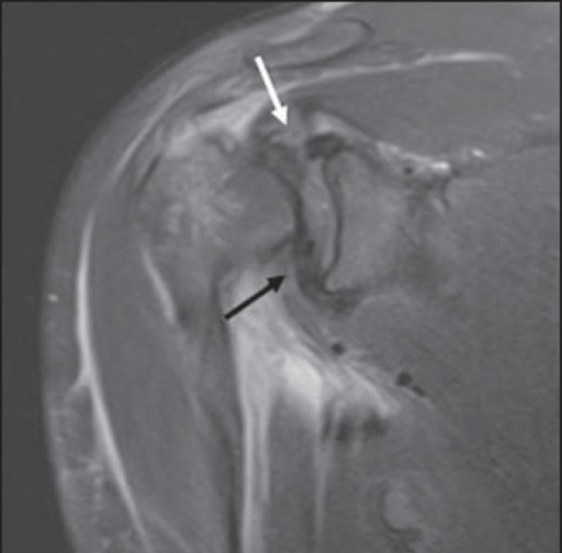
What finding is demonstrated on this coronal PD fat-saturated shoulder image?
A
Calcific tendinitis
B
Full-thickness supraspinatus tear
C
Normal supraspinatus tendon
D
Partial bursal sided tear
Click to view full size
Speed 0.75x
Speed 1x
Speed 1.25x
Speed 1.5x
Speed 2x
Q2
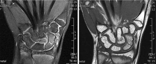
What diagnosis is shown in these images?
A
Occult scaphoid fracture
B
Lunate dislocation
C
Intraosseous ganglion
D
Normal marrow variant
Click to view full size
Speed 0.75x
Speed 1x
Speed 1.25x
Speed 1.5x
Speed 2x
Q3
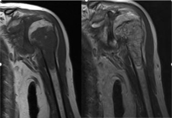
A lesion in the proximal humerus is dark on T1 and bright on T2. What does this signal pattern indicate?
A
Increased water content
B
Normal fatty marrow
C
Cortical bone
D
Air artifact
Click to view full size
Speed 0.75x
Speed 1x
Speed 1.25x
Speed 1.5x
Speed 2x
Q4
Which MRI finding is associated with a PCL tear?
A
Thickened ligament with low signal
B
Disrupted ligament with high signal
C
Intact ligament with uniform signal
D
Continuous ligament with low signal
0
of
4
answered
Submit Quiz
0%
Score
Quiz Complete!
MRI MSK Pathology
0
Correct
0
Incorrect
0
Total
Missed Questions:
0
Question Review
Retake Full Quiz
Retake Incorrect Questions Only (
0
)
Back to Free Demo
Submit quiz?
You still have unanswered questions.
Keep Answering
Submit Anyway
Question Image
Use the + / − buttons below to zoom in or out.
info@passmri.com